Carbohydrate Metabolism Disorders
Author: Alvin
Alvin
Category: Health
Carbohydrate metabolism disorders are a set of metabolic illnesses that affect the way carbohydrates are broken down. Normally, your enzymes break down carbohydrates into glucose for energy (a type of sugar). Having one of these illnesses may mean that you don’t have enough enzymes. To break down the carbohydrates in your diet. Alternatively, the enzymes may not function properly. As a result, a dangerous amount of sugar accumulates in your bloodstream. This can result in health complications, some of which are potentially life-threatening. Some of the conditions are life-threatening.
Metabolism is the process by which your body converts the energy you consume into usable energy. Proteins, carbohydrates, and fats are the building blocks of food. Enzymes, which are chemicals found in the digestive system. Beak down the dietary components into sugars and acids, which serve as fuel for your body. Your body can use this fuel straight immediately. Or it can store the energy in your body’s tissues for later consumption. A Carbohydrate Metabolism Disorders problem occurs when something goes wrong throughout this process.
These Carbohydrate Metabolism Disorders conditions are passed down through families. Many of these conditions are detected in newborn babies through blood tests. If there is a history of one of these disorders in the family. Parents can undergo genetic testing to determine whether or not they carry the gene for the disorder. Other genetic testing can be used to determine whether or not the fetus has the condition. Or contains the gene that causes it.
Special diets, supplements, and medications may be used in conjunction with other therapies. It is possible that some babies will require extra treatment if there are difficulties. Some illnesses have no known cure. But therapies may be effective in alleviating Carbohydrate Metabolism Disorders symptoms.
Glycogen storage disorders

The metabolism of the brain, red blood cells, and adrenal medulla requires a steady supply of glucose. This supply starts in the small intestine, where transport proteins help glucose into gut cells. Glucose then enters the bloodstream and is stored as glycogen in the liver. When the body is starved or fasting, glycogen is broken down into glucose. Which is subsequently released into the circulation.
Muscle tissue has glycogen stores that can be depleted by activity. Glycogen storage disorders (GSD) occur when enzymes responsible. For glycogen breakdown are hindered, causing glycogen to accumulate in the liver or muscle. These disorders can affect the liver, muscles, or both. The last stage in glucose release from the liver is faulty in GSD type I (von Gierke disease).
During infancy and early childhood, the digestive tract is continuously fed glucose (e.g., by drip feeding). Symptoms tend to improve with age. Before bedtime, eat lots of carbs and slow-release glucose (uncooked cornstarch). Liver transplantation may also be curative. But it reserved for patients who do not respond to standard therapy or develop liver cancer. The conventional treatment for the muscular variants is to avoid intense exertion.
GSD types III, IV, VI, and IX are caused by defects in earlier processes of glycogen breakdown in the liver. Lysosomal storage disorders discuss Pompe disease (GSD type II).
Gluconeogenesis is a process that produces glucose from amino acids and pyruvate. The gluconeogenic pathway includes enzymes like carboxylase and fructose-1,6-diphosphatase. These enzyme deficiencies also cause hypoglycemia, lactic acidemia, and liver enlargement. At first glance, gluconeogenesis diseases can confused with glycogen storage disorders.
Congenital Glycosylation Disorders
Congenital disorders of glycosylation. (CDG; formerly known as carbohydrate- deficient glycoprotein syndrome). These are a group of diseases that have just lately been identify. And that affect the brain as well as several other organ systems. It is the N-glycosylation pathway, which occurs in the cytoplasm and endoplasmic reticulum. Which are cellular organelles involve in the production of proteins and lipids. That is responsible for the fundamental biochemical abnormalities in CDG.
The most prevalent form of CDG caused by a deficiency. In a mannose-processing enzyme called phosphomannomutase 2. (type I). However, while some other enzymatic abnormalities have discovered. The biochemical bases of certain CDG subtypes also have not yet characterized. Typically, the typical form of CDG (type Ia) manifests itself . As poor muscle tone in infancy as well as severe developmental delay and abnormalities in the brain.
Other characteristics of type Ia children include inverted nipples and an abnormal distribution of fat. Particularly around the suprapubic region and buttocks. Hypoglycemia, seizures, stroke-like events, retinal damage, reduced cardiac contractility. Also, vomiting, liver illness, diarrhea, and a proclivity to bleed. Are some of the other characteristics of the condition. Aside from the rare type Ib disease (phosphomannose isomerase deficiency). There is no effective treatment for CDG. However, oral administration of mannose may be effective in some situations. Depending on the individual.
Fructose and Galactose Disorders
 Ph.D., MD, Assistant Professor Hanna Saturska.
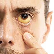
Galactosemia caused by a failure in the second major stage of galactose metabolism. Galactose-1-phosphate builds when galactose consumed. Thus, clinical signs of galactosemia start with milk feeding. The disorder causes lethargy, jaundice, liver malfunction. Also, kidney damage, and weight loss if not stopped.
Staph infections, especially Escherichia coli, can be fatal. Cataracts form if the diet contains galactose. Most galactosemic newborns have intellectual impairment if untreated or addressed late. Exclusion of galactose from the diet reverses most symptoms. Despite early therapy, most children have normal IQ. But may have learning challenges and intellectual disabilities.
Fructose-1-phosphate aldolase deficiency causes inherited fructose intolerance. HFI symptoms develop after fructose consumption. Hence they appear later than galactosemia symptoms. Fruits, sucrose, and infant formulae all include fructose. Symptoms include weight loss, vomiting, hypoglycemia, liver malfunction, and kidney disease. Older children with HFI avoid sweet foods and have teeth free of cavities. Those also with the disease fare well avoiding fructose and sugar.
Additionally, folate insufficiency impairs the ability to convert other substrates to glucose (a process called gluconeogenesis). Symptoms include severe hypoglycemia, inability to fast, and liver enlargement. The pillars of therapy are intravenous glucose and avoiding fasting. Some individuals also require nocturnal drip feedings. Or a nighttime dosage of cornstarch to treat hypoglycemia.
Lipoprotein Disorders
The principal lipoprotein classes include chylomicrons, VLDL, IDL, LDL, and HDL (HDL). Defects in lipoprotein structural proteins, cell receptors that recognize different lipoprotein types. Or enzymes that break down lipids can cause lipid metabolic disorders. As a result of these flaws, lipids can accumulate in blood vessel walls. Causing atherosclerosis. (A disease characterized by abnormal thickening and hardening of the walls of the arteries).
The lack of the LDL receptor on the surface of cells in the liver. And other organs causes familial hypercholesterolemia. Thus, cholesterol cannot enter cells. When the cell has adequate cholesterol. Feedback mechanisms instruct enzymes to stop synthesis. In familial hypercholesterolemia, these enzymes lose feedback inhibition. Resulting in increased cholesterol synthesis. It causes early coronary artery disease, strokes, and fatty deposits in the tendons. From birth, LDL cholesterol levels are higher, as well. It treated with a low-cholesterol diet and medicines that decrease cholesterol production or boost its excretion.
Severe vascular disease begins in childhood, and heart attacks are common. By the age of 20 in people with familial hypercholesterolemia. A similar phenotype seen in familial dysbetalipoproteinemia. (Hyperlipoproteinemia type III), which inherited autosomally. (That is, if the trait has inherited from both parents). An anomaly of apoprotein E, a component of lipoproteins. Causes elevated blood cholesterol and triglycerides in this adult illness. The treatment is the same as for familial hypercholesterolemia.
In abetalipoproteinemia, which is autosomal recessive, VLDL and LDL are absent. Triglycerides build up in the gut and liver, and blood levels of cholesterol. HDL cholesterol, and triglycerides are low. A lack of vitamin E causes significant fat malabsorption and neurological symptoms. Such as unstable walking, retinal abnormalities, and nerve damage.